
Anterior: thyroid, ascending aorta, brachiocephalic trunk, superior vena cava.Bifurcates at the level of T4into the left and the right main bronchus ( tracheal carina).Formed by a series of cartilages ( 15–20), joined together by annular ligaments.Laterally: pharyngeal opening of the eustachian tube, fossa of Rosenmuller, medial pterygoid plates, and superior pharyngeal constrictor muscles.Posteriorly: clivus, prevertebral musculature covering C1–C2.Contains the adenoids and eustachian tube openings.Most superior portion of the pharynx connecting the nasal cavity to the oropharynx.For more information, see “ Respiratory physiology.”.For respiratory mechanics, see pulmonary function testing.Immune defense ( ciliary clearance, alveolar macrophages, goblet cells).Respiration: gas exchange (absorption of O 2 into the blood and release of CO 2 into the air).Ventilation (distribution of air in the airway).Posterior to the lungs: vertebral column, ribs.Superior to the lungs: superior mediastinum.Between the lungs: inferior mediastinum (including heart, large vessels, tracheal bifurcation, esophagus).Lungs : located in the thoracic cavity (protected by the rib cage).Respiratory zone: lung parenchyma (site of O 2 and CO 2 exchange).Conducting zone: large airways and small airways (nonrespiratory tissue).The development of the lungs begins in the embryonic period and continues until approximately 8 years of age. The left lung shares its space with the heart, which it accommodates in the cardiac notch. The right lung consists of 3 lobes (upper, middle, lower), while the left lung consists of 2 lobes (upper, lower) and the lingula, a structure that is homologous to the middle lobe of the right lung. Gas exchange takes place in the alveoli of the lungs. Hyaline cartilage in the form of C-shaped rings ( trachea) and plates ( bronchi) provides structural protection and integrity. The entire respiratory tract down to the bronchioles is covered in ciliated epithelium, which provides immunologic protection by helping clear the airways of dust and microorganisms. The respiratory system is furthermore divided into an upper tract (structures from the larynx upwards) and a lower tract (structures below the larynx).

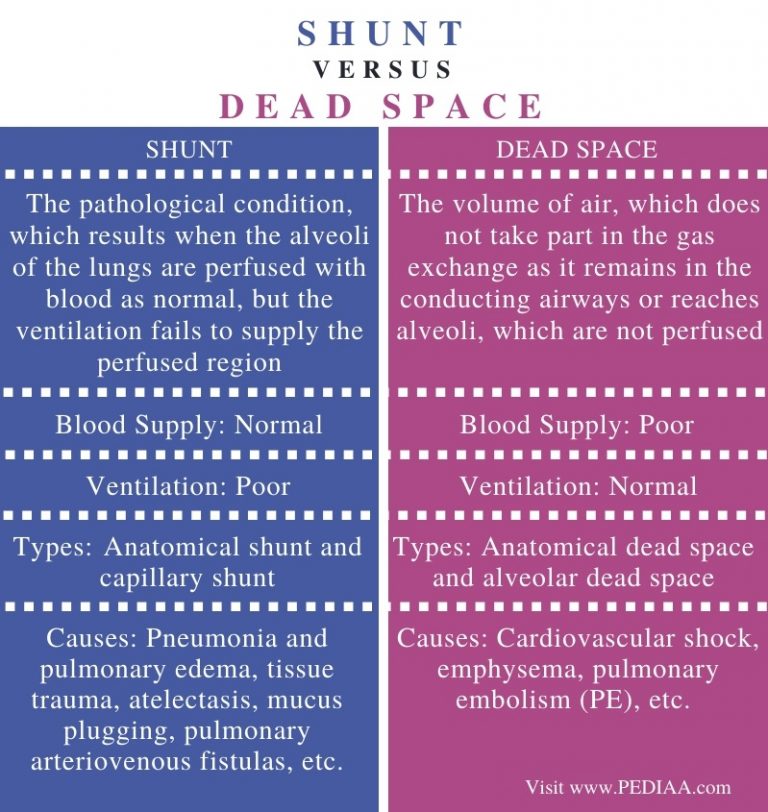
The conducting zone is composed of nonrespiratory tissue and provides the passage for ventilation of the respiratory zone, where the O 2 and CO 2 exchange takes place. The respiratory system consists of a conducting zone ( anatomic dead space i.e., the airways of the mouth, nose, pharynx, larynx, trachea, bronchi, bronchioles, and terminal bronchioles) and a respiratory zone ( lung parenchyma i.e., respiratory bronchioles, alveolar ducts, alveolar sacs).


 0 kommentar(er)
